Current Treatment Concepts for Stress Urinary Incontinence
Aktuelle Behandlungskonzepte bei Belastungsinkontinenz
Abstract
Zusammenfassung. Eine Belastungsinkontinenz sollte immer zuerst konservativ behandelt werden. Schon eine Gewichtsreduktion, Hormonpräparate, Physiotherapie, Beckenbodentraining und/oder die Anwendung von Pessaren können zum Erfolg führen. Nach Ausschöpfen dieser Therapien werden heute Inkontinenzoperationen mit meist sehr guten Heilungschancen (ca. 80–90 %) angeboten. Der operative Goldstandard ist die suburethrale Schlingeneinlage. Die Pelvic-Floor-Sonografie liefert dazu sehr wichtige Hinweise zur Wahl der Operationstechnik und zur Behebung von Komplikationen. Ferner bildet die intra- oder paraurethrale Injektion von Bulking Agents eine vielversprechende, wenig invasive operative Alternative. In diesem Artikel werden Behandlungskonzepte, prä-, intra- und postoperative Untersuchungen, Wahl der Operationsmethode, operationstechnische Details für den Operationserfolg sowie Vorbeugung und Behandlung von Komplikationen diskutiert.
Abstract. Initially, stress urinary incontinence should be treated by conservative measures, such as weight reduction, hormonal substitution, physiotherapy, pelvic floor exercise and/or the use of pessaries. Incontinence surgeries are only recommended in case of unsuccessful conservative therapy. Today, tension-free suburethral sling insertions represent the gold standard of incontinence surgery yielding very good outcomes (cure rates of 80–90 %). Pelvic-floor sonography provides important information on decision of surgical methods and the management of complications. Furthermore, intra- or paraurethral injection of bulking agents is a promising, minimally invasive surgical alternative. This article discusses treatment concepts, pre-, intra- and post-operative examinations, decision on surgical methods, operational details for surgical success, and the prevention and management of complications.
Résumé. En première intention, l’incontinence urinaire doit être traitée par des mesures non invasives, telles que la réduction de poids, la substitution hormonale, la physiothérapie, l’exercice du plancher pelvien et/ou l’utilisation de pessaires. Après avoir épuisé toutes ces thérapies, la chirurgie de l’incontinence offre de très bons résultats. Aujourd’hui, la bandelette synthétique sous urétrale représente l’intervention de référence (ou gold standard) de la chirurgie de l’incontinence avec un taux de guérison de 80–90 %. L’échographie pelvienne fournit des informations importantes sur la décision des méthodes chirurgicales et la prise en charge des complications. En outre, l’injection d’agents comblants intra- ou para-urétrale offre une alternative chirurgicale prometteuse et moins invasive. Cet article discutera des concepts de traitement, des examens pré-, per- et post-opératoires, du choix des méthodes chirurgicales, des détails techniques pour la réussite chirurgicale, et de la prévention et du traitement des complications.
Introduction
Urinary incontinence is defined as an involuntary loss of urine. It is not an independent disease, but a symptom of various etiologies with different disease severities [1]. Urinary incontinence often leads to social exclusion and a significant restriction in quality of life. In Switzerland, approximately 400,000 women are affected [2]. Stress urinary incontinence representing 60 % of all cases is the most frequent form of incontinence [3]. Five to 20 % of all women are affected [4] whereby this percentage continuously increases with age.
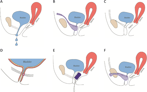
Stress urinary incontinence is characterized by an involuntary loss of urine due to physical activities, with an increased intra-abdominal pressure, but without an urodynamically diagnosed detrusor instability [5, 6, 7]. In this case, the urethral closure mechanism, effective by the urethral closure pressure at rest and the pressure transmission during physical activity, cannot withstand the actual increase of intra-abdominal pressure [8]. Depending on the severity of stress urinary incontinence during physical straining, leakage may be as little as a drop or two, or may be a “squirt,” or even a stream of urine (Fig. 1-A).
Numerous studies [9, 10] confirmed the integral theory by Ulmsten and Petros [11] that suggested causes for the decrease in urethral closure pressure. These six anatomical structures are likely to be involved: Dysfunction in the region of bladder neck or striated muscles, loosening of the ligamenta sacrouterina and the attachment site of the pubococcygeus muscle, insufficiency of the pubourethralia ligament and the suburethrally situated vaginal wall. According to Ulmsten and Petros, the two last-mentioned structures are most important for correct functioning. Factors that lead to weakening of muscle and connective tissue in the pelvic floor area are increasing atrophy of the urethral mucosa after postmenopausal estrogen deficiency, a decrease in vascularization, pregnancy, childbirth, obesity, and a severe intra-abdominal pressure increase by chronic cough or constipation [1, 12].
The integral theory provided the base for the development of the suburethral sling insertion technique. In the mid-1990s, this new operative approach revolutionized incontinence surgery [13]. Continence was achieved by the tension-free support of the midurethra by a synthetic polypropylene tape (TVT) [14, 15]. Today, this method is the gold standard for the treatment of stress urinary incontinence (Fig. 1-B).
Conversely, the more invasive colposuspension technique (Fig. 1-C), the former gold standard, is rarely used, and typically, in combination with a laparoscopic prolapse surgery. In general, prolapse and incontinence surgeries are not combined, but performed consecutively with the prolapse surgery preceding the tape insertion [16]. Today, the intra- or paraurethral injection of bulking agents, such as Bulkamid®, offers a further treatment alternative to TVT insertion (Fig. 1-D).
Conservative therapy of stress urinary incontinence
Guidelines recommend to fully exploiting conservative measures for the treatment of stress urinary incontinence before considering operative therapies [17]. Conservative measures include weight reduction and the local application of hormones for epithelial proliferation. Products with estriol (E3) are preferred over products with estradiol (E2). Physiotherapy is another important treatment element. If necessary, it can be used in combination with electrostimulation, biofeedback or whole body vibration therapy [18] to strengthen the pelvic floor muscles and to optimize the muscular coordination. Furthermore, pessaries (ring or bowl shaped, made of silicone, disposable pessaries) are used for urethral support (Fig. 1-E). They are inserted with the help of an estrogen cream (E3), and can be changed daily and independently by the patient.
If all these conservative measures are unsatisfactory, an operative therapy needs to be considered.
Operative therapy
Tension-free tape insertion
Preoperative investigation
Before deciding for surgery, all conservative treatment options should be exhausted, the tissue should be well-estrogenized and built-up and thus, suitable for the operative intervention. In addition to clinical examination and urodynamics, pelvic floor sonography is very important for the precise planning of the incontinence surgery. Pelvic floor sonography enables the assessment of the entire small pelvis including the anterior, apical and posterior compartments [19].
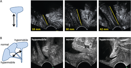
Sonographic findings allow very precise recommendations for surgical indications and the acquisition of operationally important details to plan the intervention. According to radiological and morphological investigations by Ulmsten and Petros, the suburethral sling has to be positioned precisely within the middle third of the urethra, the key area for achieving continence, between the point of maximal urethral closure pressure at rest and the urethral knee which anatomically corresponds to the attachment site of the ligamenta pubourethralia [11, 20, 21]. Our studies [22, 23, 24, 25, 26] published in recent years showed that a detailed sonographic diagnosis of the lower urinary tract with special consideration of the physiologically variable urethral length allowed an exact positioning of the sling and thus, optimizes surgical outcome. Assessments of urethral length and mobility (Fig. 2) and the height of the paraurethral sulci (Fig. 3) are crucial for selecting the best suited pathway (retropubic or transobturator approach) and the optimal placement of the suburethral sling.
Types of tapes
Incontinence surgeries with tension-free tapes are performed under local anesthesia with analgosedation. Thereby, a macroporous monofilament band of polypropylene is suburethrally inserted. Fibroblasts eventually migrate into the band resulting in a “natural reconstruction” of the tissue. This leads to a new support of the urethra at stress condition.
The suburethral slings currently available on the market require different insertion and fixation techniques. They can be divided into three groups.
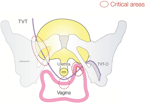
The placement of the classic TVT is retropubic (Fig. 1-B), while the placement of the TOT is by the transobturator approach (Fig. 1-F). Both options are equally successful regarding cure rates of stress incontinence (80–95 %) [17, 27]. Regarding complications, bladder injuries and emptying disorders are more frequent with the retropubic method, while pain (hip/groin/thigh/pubic bone), dyspareunia and band erosions (Fig. 3) are more frequent with the transobturator method. In case of an elevated proximal urethra and a high, well preserved sulcus vaginalis (nullipara/status after preoperations), the classic retropubic tape has a better outcome than the transobturator tape [17]. In case of a low position of the urethra and a flat sulcus vaginalis (middle-sized cysto urethrocele/rotatory descent; no preoperations) also a transobturator placement (TOT) can be considered. Furthermore, it has to be taken into account that retropubic tapes tend to pull towards the proximal, and transoburator tapes towards the distal direction [26, 28].
The third innovation involves slings with short arms. With this alternative, the operation procedure should be less invasive and the risk of injury and pain should be reduced. But these so-called “mini slings” are no valuable alternative to the classic TVT. First, long-term records are missing and second, initial data suggest a suboptimal stabilization of the tape and lower cure rates [17]. In addition, the two most commonly used mini slings are no longer available on the market.
Intraoperative assessments
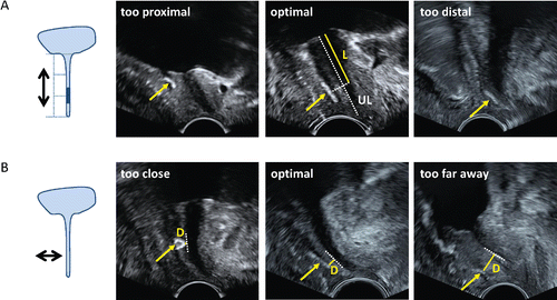
The incision site should intraoperatively be determined based on the sonographically measured urethral length (Fig. 2-A). Ideally, the optimal tape position is at the transition of the middle to the distal third of the urethra (Fig. 4-A). This can be achieved by starting the incision at a distance from the external urethral orifice corresponding to one-third of the sonographically determined urethral length, i. e., an urethral length of 45 mm requires an incision at 15 mm distance, and a short urethral length of 18 mm requires an incision starting at 6 mm from the external urethral orifice.
Furthermore, the evaluation of the urethral mobility is important since the tension-free positioning of the tape also depends on this parameter (Fig. 2-B). For a rotatory descensus urethrae and a normal and hypermobile urethra, the straightforward intraoperative determination of the tape-urethra distance with the Cooper scissors is sufficient to achieve a tension-free position of the tape. However, for a vertical descensus and a hypo- and immobile urethra, this procedure can lead to a too large tape-urethra distance and to only a minimal improvement of the incontinence, or even to therapeutic failure. In these cases, the intra-operative cough test is very important to adjust the tape with the low mobility of the urethra.
A correct operative positioning of the tape is more successful under physiological anatomical conditions (normal, non-relaxed tone of the pelvic floor). This requires an operative procedure in local anesthesia under analgosedation, as initially described [12]. Prerequisites are: An exact operative technique (precise setting of local anesthesia), a careful operation and a well-coordinated timing of operative steps and anesthesia (sedation/setting of local anesthesia/incision and pull through of the tape/cystoscopy/ cough test).
The distance between tape and urethra has a large impact on success and complication rates of the operation (Fig. 4-B). A large tape-urethra distance of >5 mm is associated with a lower cure rate. Patients with a too closely positioned tape, i.e. at <3 mm from the urethra, are significantly more often confronted with obstruction complications like voiding dysfunction and/or de novo urge incontinence. The tape should be located at the transition site of the middle to the distal third of the urethra (Fig. 4-A), with an ideal tape-urethra distance between 3 and 5 mm (Fig. 4-B), and without urethral kinking.
Recovery time after this operation is short and surgical scars are minimal. Long-term data show an objective cure rate of 83 %, and a high subjective satisfaction rate of up to 95 % [23].
Postoperative evaluation and complications after tape insertion
Frequency and severity of complications after incontinence surgeries [17] depend, inter alia, on the operative technique (type of tape, following a standardized procedure) and on the experience of the surgeon. Early complications are bladder perforations, bleedings in the puncture channel, urinary retention and persistent stress urinary incontinence. Late complications, which may develop after years and become increasingly disturbing, are micturition disorders, urge and pain symptoms, residual urine formation, recurrent urinary tract infections, irritable bladder problems, ulcer formation in the area of the tape, dyspareunia and also recurrent stress urinary incontinence.
It is important to identify an incorrectly positioned (dystopic) tape and to adjust it as soon as possible [25]. The early postoperative urogynecologic pelvic floor sonography proves to be essential to detect the so-called “failures” of the method or to find the causes for postoperative complications. Pelvic floor sonography allows a good visualization of the tape position relative to the urethra (Fig. 4). Furthermore, tape-related, obstructive micturition disorders with residual urine can easily be detected at an early stage. Postoperative seromas and hematomas can also be identified. Therefore, in presence of increased postoperative residual urine values or symptoms of urge/pain [29], an ultrasound analysis in sagittal (Fig. 4) and transversal planes should be performed in the first postoperative days.
After TVT insertion, voiding dysfunction can be observed for 5 % of the patients. Very often, this complication is caused by a dystopic tape position (tape too tight, too close to the urethra or at the level of the bladder neck). But voiding dysfunction can also be caused by tape distortion, by only a punctual action of the tape on the urethra, or by kinking of the urethra.
Tape mobilization and tape incision
Tape mobilization is a simple and successful method to cure urethral obstruction with disturbed voiding due to a too tightly positioned or C-shaped tape, without compromising the outcome of the original stress urinary incontinence surgery [25]. This method is performed by reopening the suburethral incision within the first days after tape insertion (Fig. 5) [25, 30]. In cases of an asymmetrically placed tape, pelvic floor sonography also allows to localize the ideal side for tape loosening. Tape mobilization usually is not necessary for tape-urethra distances >3 mm, since in most cases, voiding dysfunctions resolved simultaneously with reduction of edema, pain and muscle contractions and with resorption of the hematomas. However, smaller tape-urethra distances (<3 mm) require a tape mobilization. Also here, the experience of the surgeon plays an important role. When the tape is only stretched, but not actually mobilized, it simply returns back into its original position. Conversely, overly aggressive tape loosening increases the risk for recurrent stress urinary incontinence. Price et al. [31, 32] identified the benefits of tape mobilization immediately after TVT placement as a resolution of voiding problems and to avoid long-term catheterization.
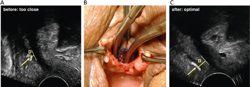
Already a few days after tape insertion, fibroblasts grow into the polypropylene band. Then, urinary retention can only be cured by sling incision. However, this procedure results in a high recurrence rate of stress urinary incontinence of up to 50 % [32]. In cases of voiding dysfunction, or symptoms of urge and urethral pain, the sling is dissected suburethrally, a slightly opened clamp is placed below it and the sling is completely severed with scissors in between the arms of the clamp. In presence of mesh exposure, the visible part of the sling needs to be resected underneath the healthy skin, sufficiently distant from the exposure.
In contrast to tape incision, early tape mobilization represents a much simpler and more successful method to treat voiding dysfunction due to a too tightly positioned tape. Furthermore, tape mobilization can resolve this frequent complication without requiring a second hospitalization and without generating much additional costs. Unfortunately, the resolution of simple complications often starts too late [33] and is nonprofessional (extensive conservative therapy instead of surgery, or only incomplete tape incision). Frequently, this results in tedious, unnecessary suffering. If stress urinary incontinence persisted after the first tape insertion, a second tape can be placed, provided that the urodynamic assessment shows a functional-operative indication.
Colposuspension
Before the suburethral sling procedures established as the gold standard, Burch colposuspension in the modification of Cowan was the standard therapy for stress urinary incontinence [34]. This method includes the loose approximation of the lateral edges of the vagina to Cooper’s ligament by using non-absorbable sutures (Fig. 1-C). This results in a hammock-like suspension of the urethra and a fixation of the bladder neck [34, 35]. Cure rates of 70–90 % are similarly high as for sling insertions [36]. However, the procedure of colposuspension is much more complex and invasive, and requires an abdominal open or laparoscopic surgery with general anesthesia. Therefore, colposuspension is rarely used in primary therapy, and limited to exceptional cases such as the presence of a hypermobile urethra, or the concomitant prolapse surgery. Postoperative complications of colposuspension are voiding dysfunction due to overcorrection, de novo urgency/ urge incontinence symptoms, and rectocele and enterocele [37].
Bulking agents
Alternatively, stress urinary incontinence can also be treated by intra- or paraurethral injection of bulking agents (Fig. 1-D). Deposits of bulking agents lead to a constriction (coaptation) of the urethra. Generally, this procedure only requires a local anesthesia, and hence, represents one of the least invasive operative therapies of incontinence.
Since the 1970s, a number of bulking materials have been tested, e. g. teflon, autologous fat, bovine or porcine collagen, silicone particles, dextranomer/hyaluronic acid copolymer, ethylene vinyl alcohol copolymer, calcium hydroxylapatite, polyacrylamide hydrogel and carbon beads [38, 39]. Most materials have been withdrawn from the market due to different reasons. Currently, there are three injectable bulking agents approved by the Food and Drug Administration (FDA) available on the market in the United States: Durasphere® (carbon-coated beads), Coaptite® (calcium hydroxylapatite particles in a gel carrier) and Macroplastique® (polydimethylsiloxane) [40]. Bulkamid®, a polyacrylamide hydrogel (PAHG), is available on the European market. It is the most frequently used bulking agent in Europe, has very good qualities, and data of several years of experience are available [41]. It is sterile, non-odorous and transparent, and made up of 2.5% cross-linked polyacrylamide (dry weight) with 97.5% water. PAHG is biocompatible, biologically non-degradable, not absorbable, migration resistant, not toxic and not allergenic [39]. Under cystoscopic control, three to four small submucosal deposits of the PAHG filling substance are placed transurethrally at the transition of the proximal urethra to the midurethra. This leads to the support and the constriction of the urethra.
Bulking agent therapy can be used as primary or secondary treatment of stress urinary incontinence. Primary injection therapy is recommended for patients with complicating concomitant findings, such as pronounced obesity, comorbidities, complications with general anesthesia, vast varicosis in the small pelvis, and status after radical operation of cervix or endometrium carcinoma, or after radiation therapy. Further indications for bulking agent therapy are requests for a minimally invasive procedure or for a mesh-free alternative. Secondary injection therapy is suited for patients after failed previous incontinence surgeries such as colposuspension or sling insertion [42, 43]. Studies of bulking agent therapy after failed midurethral sling insertion are rarely described in literature, and can only be found in a retrospective context [42, 44]. Bulking agent injections offer great advantages for patients with concomitant urge symptoms or with an immobile urethra due to a preceding surgery (e.g. colposuspension). The method is easy to perform and has low complication and high cure rates, particularly in cases of high-risk patients. Risks factors should individually be evaluated, especially for patients with recurrent stress urinary incontinence after sling failure. In presence of the above mentioned risk factors, bulking agent injection can be offered. It will be interesting to see the results of a currently ongoing prospective randomized Finnish study that directly compares TVT and Bulkamid® therapy for the indication of primary stress urinary incontinence. Patient satisfaction, complication rates, and improvement of continence will be evaluated (study number NCT02538991, https://clinicaltrials.gov/).
Conclusions
Suburethral sling insertion represents the current operative gold standard to treat stress urinary incontinence. A preoperative sonographic assessment should evaluate the patient’s individual characteristics for the optimal positioning of the tape. Furthermore, a good postoperative management of complications will minimize long-term suffering. Postoperative voiding dysfunctions can be cured by tape mobilization within the first few days after sling insertion. In contrast to tape incision, early tape mobilization does not impair the chances of cure of the incontinence surgery. Furthermore, intra- or paraurethral injection of bulking agents is a promising minimally invasive alternative to treat stress urinary incontinence.
Key messages
- •Primarily, stress urinary incontinence should be treated by conservative measures.
- •Operative options are: suburethral sling insertions (retropubic or transobturator, mini slings), or the intra- or paraurethral injection of bulking agents.
- •A good build-up of the tissue for surgery, the experience of the surgeon, the consideration of individual characteristics, and an effective management of complications all contribute to the success of the incontinence surgery.
- •Pelvic floor sonography is crucial for planning, performing and evaluation of incontinence surgeries.
Questions
1. Which therapy options are used for the conservative treatment of stress urinary incontinence? (2 correct answers)
- a)Use of pessaries
- b)Anticholinergic agents
- c)Local applications of hormones
- d)Colposuspension
2. Today, which is the most frequently used surgical method to treat stress urinary incontinence? (1 correct answer)
- a)Colporrhaphia anterior
- b)Suburethral sling insertion
- c)Colposuspension
- d)Paraurethral injection of bulking agents
Answers
1. Answers a) and c) are correct.
Anticholinergic agents are used to conservatively treat urinary urge incontinence. Colposuspension is an operative and not a conservative therapy.
2. Answer b) is correct.
TVT insertion or suburethral sling insertion currently is the most frequently used operative therapy to cure stress urinary incontinence.
Abbreviations:
E2: Estradiol
E3 Estriol
PAHG Polyacrylamide hydrogel
TVT Retropubic tension-free vaginal tape
TOT Transobturator vaginal tape
Literature
: Female incontinence: work-up and therapy. Praxis 1995; 84: 726–735.
: Die Belastungs-inkontinenz. Diagnostik und Therapie. Gynäkologie 2013; 5: 12–17.
. Urogynäkologische Anamnese. In: Tunn R, Hanzal E, Perucchini D (Hrsg.). Urogynäkologie in Praxis und Klinik. Berlin; De Gruyter: 2009. 59–68.
, : Understanding the burden of stress urinary incontinence in Europe: a qualitative review of the literature. Eur Urol 2004; 46: 15–27.
, : An International Urogynecological Association (IUGA)/International Continence Society (ICS) joint report on the terminology for female pelvic floor dysfunction. Neurourol Urodyn 2010; 29: 4–20.
, : The standardisation of terminology in lower urinary tract function: report from the standardisation sub-committee of the International Continence Society. Urology 2003; 61: 37–49.
, : Fourth International Consultation on Incontinence Recommendations of the International Scientific Committee: Evaluation and treatment of urinary incontinence, pelvic organ prolapse, and fecal incontinence. Neurourol Urodyn 2010; 29: 213–240.
: Gynecologic urology. Gynakol Rundsch 1991; 31(Suppl 1): 1–52.
: An integral theory of female urinary incontinence. Experimental and clinical considerations. Acta Obstet Gynecol Scand Suppl 1990; 153: 7–31.
, : Paraurethral connective tissue in stress-incontinent women after menopause. Acta Obstet Gynecol Scand 1998; 77: 95–100.
: Different biochemical composition of connective tissue in continent and stress incontinent women. Acta Obstet Gynecol Scand 1987; 66: 455–457.
: Some reflections and hypotheses on the pathophysiology of female urinary incontinence. Acta Obstet Gynecol Scand Suppl 1997; 166: 3–8.
: An ambulatory surgical procedure under local anesthesia for treatment of female urinary incontinence. Int Urogynecol J Pelvic Floor Dysfunct 1996; 7: 81–85; discussion 85–86.
: Vaginal mesh grafts and the Food and Drug Administration. Int Urogynecol J 2010; 21: 1181–1183.
: Classification of biomaterials and their related complications in abdominal wall hernia surgery. Hernia 1997; 1: 15–21.
: Prolapse surgery with or without stress incontinence surgery for pelvic organ prolapse: a systematic review and meta-analysis of randomised trials. BJOG 2014; 121: 537–547.
, : Interdisciplinary S2e Guideline for the Diagnosis and Treatment of Stress Urinary Incontinence in Women: Short version – AWMF Registry No. 015–005, July 2013. Geburtshilfe Frauenheilkd 2013; 73: 899–903.
, : Pelvic floor muscle training for stress urinary incontinence: a randomized, controlled trial comparing different conservative therapies. Physiother Res Int 2011; 16: 133–140.
: Belastungsinkontinenz – Individuell behandeln dank optimaler Diagnose. J Urol Urogynäkol 2010; 17: 50–52.
: Anatomical assessment of the bladder outlet and proximal urethra using ultrasound and videocystourethrography. Int Urogynecol J Pelvic Floor Dysfunct 1998; 9: 365–369.
: An integral theory and its method for the diagnosis and management of female urinary incontinence. Scand J Urol Nephrol Suppl 1993; 153: 1–93.
, : Can we place tension-free vaginal tape where it should be? The one-third rule. Ultrasound Obstet Gynecol 2012; 39: 210–214.
, : Tape functionality: sonographic tape characteristics and outcome after TVT incontinence surgery. Neurourol Urodyn 2008; 27: 485–490.
: Introital ultrasound in the diagnosis of occult abscesses following a tape procedure: a case report. Arch Gynecol Obstet 2013; 288: 577–579.
, : Ultrasound and early tape mobilization – a practical solution for treating postoperative voiding dysfunction. Neurourol Urodyn 2014; 33: 1147–1151.
, : Do different vaginal tapes need different suburethral incisions? The one-half rule. Neurourol Urodyn 2015; 34: 741–746.
: Transobturator tapes: better than TVT?Gynakol Geburtshilfliche Rundsch 2006; 46: 79–82.
, : Tape functionality: position, change in shape, and outcome after TVT procedure – mid-term results. Int Urogynecol J 2010; 21: 795–800.
: Urgency after a sling: review of the management. Curr Urol Rep 2014; 15: 400.
: The effect of time to release of an obstructing synthetic mid-urethral sling on repeat surgery for stress urinary incontinence. Neurourol Urodyn 2017; 26: 349–353.
: The benefit of early mobilisation of tension-free vaginal tape in the treatment of post-operative voiding dysfunction. Int Urogynecol J Pelvic Floor Dysfunct 2009; 20: 855–858.
, : Midurethral sling incision: indications and outcomes. Int Urogynecol J 2013; 24: 645–653.
, : Reasons for and treatment of surgical complications with alloplastic slings. Int Urogynecol J Pelvic Floor Dysfunct 2006; 17: 3–13.
. Gynäkologische Urologie: Interdisziplinäre Diagnostik und Therapie. 4. Auflage. Stuttgart; Thieme: 2013.
: Vaginale Inkontinenzioperationen – wann, wie und warum?J Urol Urogynäkol 1999; 1: 34–38.
: Open retropubic colposuspension for urinary incontinence in women. Cochrane Database Syst Rev 2016; 2: CD002912.
, : Introital ultrasound of the lower genital tract before and after colposuspension: a 4-year objective follow-up. Ultrasound Obstet Gynecol 2004; 23: 277–283.
, : Urethral injection therapy for urinary incontinence in women. Cochrane Database Syst Rev 2012: CD003881.
. Harninkontinenz der Frau: Bulking-Agents als moderne Therapieoption. Bremen; UNI-MED: 2012.
: Update on urethral bulking agents for female stress urinary incontinence due to intrinsic sphincter deficiency. J Urol Res 2014; 1: 1009.
: Long-term effectiveness and durability of Bulkamid® as primary treatment of stress urinary incontinence – a longitudinal study. Presented at ICS 2015, Montreal, Canada, abstract 356.
: Urethral bulking for recurrent stress urinary incontinence after midurethral sling failure. Neurourol Urodyn 2017; 36: 722–726.
: Transurethrale Injektionen mit Bulkamid. Geburtshilfe Frauenheilkd 2010; 70: 47–51.
: Repeat midurethral sling compared with urethral bulking for recurrent stress urinary incontinence. Obstet Gynecol 2014; 123: 1207–1212.


